Sleep / Editorial
What happens to our breathing during sleep?
Introductory · 8 min read · ■■■□ moderate evidence
During the day we can hold our breath or speed it up if we want. Once we fall asleep, the brain takes over completely. All night, without our noticing, we keep breathing in and out. Most nights this happens quietly. On some nights, though, something changes in the throat: snoring starts first, and sometimes breathing stops altogether for a while.
In this article we explain from the beginning what happens to breathing during sleep, which of it is harmless, and what deserves attention.
Breathing through the night
Sleep is not one long stretch. A night is made of cycles that each last 80 to 100 minutes, and there are usually four to six of them1. In each cycle we move from light sleep into deep sleep, then into REM sleep, when most dreams happen. In REM the muscles go slack and stay still, and this stage gets longer towards morning1.
It is not only the arms and legs that relax. As we fall asleep, the muscle in the tongue relaxes too: in one study of this muscle, about half of the nerve cells that fire with each breath fell completely silent at the moment of falling asleep7. This is why, in some people, breathing problems are worst in REM sleep, in the early hours of the morning8.
The three states of the throat
Part of the passage from the nose to the lungs is not surrounded by bone: the soft palate, the base of the tongue and the walls of the pharynx are flexible tissue (Figure 1).
- Nasal cavity. Most breathing comes in here. The air is warmed and moistened on the way.
- Hard palate. The bony front part of the roof of the mouth. It does not move during sleep.
- Soft palate. The flexible, muscular part behind the hard palate, with the uvula at its tip. One of the tissues that vibrates in snoring.
- Tongue. A large mass of muscle. Like the other muscles, it relaxes during sleep.
- Tongue base. The back of the tongue, facing the throat. It can slide backwards when you lie on your back and the muscles relax.
- Pharynx. The passage for air coming from the nose and mouth. This is where the airway narrows during sleep.
- Back wall of the pharynx. The wall in front of the spine. When the airway closes, the soft palate and tongue base rest against it.
- Epiglottis. The flap that covers the entrance to the windpipe when you swallow.
- Hyoid bone. A small bone that the tongue and throat muscles attach to.

When the muscles relax, this soft section narrows a little2. The narrowing has three states (Figure 2):
- Airway open. Air passes quietly. This is quiet breathing.
- Airway narrowed. Air rushing through the narrow gap makes the soft tissues vibrate. The sound of that vibration is snoring.
- Airway closed. No air gets through. The chest and belly keep trying to breathe, but no sound comes from the mouth or nose. This is called apnea. The brain restarts the muscles with a brief awakening; that deep, rasping first breath is the moment it happens2.
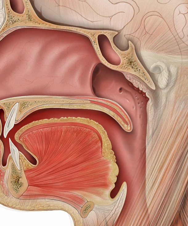 Airway openAir passes quietly.
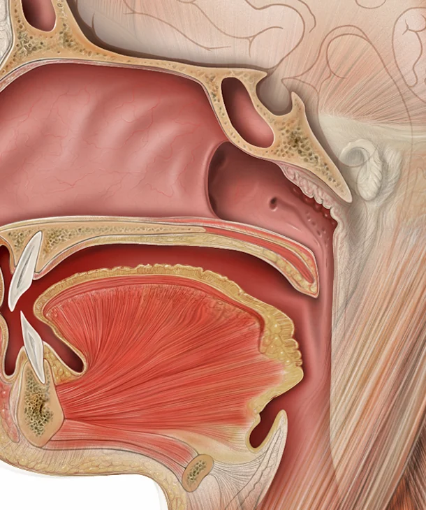 Airway narrowedThe tongue base and soft palate slide back; air rushing through the narrow gap makes the tissues vibrate.
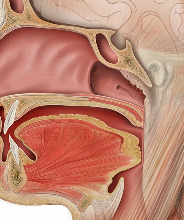 Airway closedThe soft palate and tongue base rest against the back wall; no air passes and the sound stops.
These awakenings are usually not remembered in the morning. The person thinks they slept through the night, but their sleep was broken up again and again.
Snoring does not mean being ill
Snoring is very common. In a nationwide study of 5,021 adults from every region of Türkiye, one in ten (9.6%) said they snored almost every night3. In most of these people, breathing does not stop. The classification used by sleep physicians does not count snoring on its own as a disease either; it lists it among the "normal variants"4.
So there is no need to panic because the person next to you snores. What is worth listening to is not the sound itself, but the gaps in it.
The real sign: silence
It is three in the morning. The person beside you is snoring; a steady sound you have been used to for years. Then the sound suddenly stops. The room is quiet. One, two, three seconds. Five. Ten. Then a deep, rasping breath, and the snoring carries on where it left off.
Most people do not notice that silence, and those who do usually think "well, it passed". Yet this may be the moment the airway closed completely. In sleep apnea the airway closes tens, sometimes hundreds of times a night. Doctors measure it by how many times it happens per hour. Worldwide, about 936 million people aged 30 to 69 are estimated to have mild or more severe sleep apnea, and 425 million of them moderate or severe sleep apnea9. In the study from Türkiye, 13.7% of adults came out as high risk for sleep-disordered breathing3.
Most people do not know this, because they cannot hear their own sleep. A study in the United States estimated that 93% of women and 82% of men with moderate or severe sleep apnea had not been diagnosed10. These figures are from the 1990s10; we could not find a newer large study that answers the same question today. But the core of the problem has not changed: apnea is usually noticed not by the person sleeping, but by the person next to them.
Not every silence is apnea. The person may have turned over, closed their mouth or settled into quiet breathing. That is why a single night of listening is not enough to decide. What matters are long, repeated silences: longer than ten seconds, ending in a deep gasp, and coming back again and again through the night.
What can sound tell, and what can it not?
A phone microphone hears snoring well. In a study that tested three snoring apps in a sleep laboratory, the snoring the apps counted agreed with what the laboratory counted11. A model trained on home recordings estimated the share of the night spent snoring almost exactly as the experts had marked it12.
Telling from sound alone whether breathing has stopped is harder. A pooled analysis of eleven studies on phone-based sleep apnea screening found that methods relying on sound alone caught most people with apnea, but were less good than methods measuring movement and oxygen at telling apart people without apnea5. An international expert group also did not consider sound alone enough to grade how serious snoring is13.
In short, sound is a good clue, not a diagnosis. Sleep apnea is diagnosed with a sleep test, done in a laboratory or at home6.
When to see a doctor?
If someone has seen your breathing stop in your sleep, if you nod off while driving, or if you wake up feeling like you are choking.
If you wake up with headaches, feel tired all day, or have high blood pressure. The sleep centres accredited by the Turkish Sleep Medicine Society are listed by province.
We have gathered what you can bring and what you can ask when you see a doctor on a separate page.
What can you do tonight?
If someone next to you snores, listen to the silences and make a note: roughly how many seconds they lasted, how many times they happened in the night, whether the person was lying on their back, and whether each one ended with a deep gasp. When you talk to a doctor, these few lines say far more than "they snore a lot".
If you are the one snoring, look at what makes it worse. Sleeping on your back, alcohol and late meals in the evening, weight and a blocked nose can all make snoring worse. We also look at which remedies work and which are only popular on social media, with sources, in the Myth or fact? column.
If you are curious, you can listen to clips from real sleep laboratory recordings to hear what snoring and pauses in breathing sound like.
In depthmechanisms, studies, limitations
Pathophysiology. Obstructive sleep apnea does not have a single cause. A narrow or easily collapsible upper airway is the basic predisposition; it is accompanied, in varying proportions, by an insufficient dilator muscle response during sleep, a low arousal threshold and an oversensitive ventilatory control (high loop gain)2. Snoring is the vibration of the soft palate, uvula and tongue base in the partly collapsed segment; most events end with a cortical arousal, and sleep becomes fragmented2.
Site of collapse. Collapse can occur behind the soft palate, behind the tongue base, or at both; its site and pattern differ between people2. Below is the sound of the same three states, recorded above the bed in a sleep laboratory. In the spectrogram each breath shows up as a vertical band; in apnea the bands stop within the intervals marked by the expert.
ABC- A. Retropalatal region: velopharynx; between the velum palatinum and the posterior pharyngeal wall.
- B. Retroglossal region: oropharynx; between the radix linguae and the posterior pharyngeal wall.
- C. Epiglottis: the flap over the larynx.
Airway open: quiet breathing

Airway narrowed: regular snoring

Airway closed: recurrent obstructive apnea

Definition and thresholds. According to ICSD-3-TR, a diagnosis of OSA requires at least 5 predominantly obstructive respiratory events per hour on PSG or HSAT together with symptoms or an associated condition, or at least 15 events per hour regardless of symptoms4. Under the AASM rule, a hypopnea is scored as a 30% reduction in airflow accompanied by a 3% desaturation or an arousal14; in centres using the 4% rule, the AHI of the same night comes out lower, and this difference is not evenly spread across age groups15.
Prevalence. In the Swiss HypnoLaus cohort (aged 40 to 85), an AHI of 15 or more was found in 49.7% of men and 23.4% of women16. A literature-based global estimate projects 936 million mild or worse and 425 million moderate or severe cases between the ages of 30 and 699. In TAPES (Türkiye, n=5,021), high risk on the Berlin questionnaire was 13.7% and habitual snoring 9.6%; because it is questionnaire based, this is not PSG-confirmed prevalence3.
Performance of acoustic methods. In a meta-analysis of eleven studies (AHI 15 or more), the pooled sensitivity of phone-based methods was 0.91 and the specificity 0.88. In the sound-only subgroup (5 studies), sensitivity held at 0.88 while specificity fell to 0.81; in the movement-based subgroup specificity was 0.965. A deep learning model tested per person on home recordings reported r=0.97 agreement for the snoring ratio12. In commercial apps, agreement between snore counts and PSG is good, while the relationship with AHI is moderate and varies by app11. There is no consensus on the definition, thresholds and microphone position for objectively measuring snoring17.
Guideline. The AASM diagnostic guideline recommends that questionnaires and clinical prediction tools not be used to diagnose OSA without PSG or HSAT. In a patient with excessive daytime sleepiness plus at least two of habitual loud snoring, witnessed apnea or gasping or choking, and diagnosed hypertension, the likelihood of moderate to severe OSA is high; such a patient should have PSG or a technically adequate HSAT6.
Limitations. The most often cited estimate of the share of undiagnosed disease dates from 1997 and comes from a sample of employed adults in the United States10; there is no current, population-based, PSG-confirmed counterpart. We could not find national prevalence data for Türkiye measured with PSG.
Sources
- National Heart, Lung, and Blood Institute. How Sleep Works: Sleep Phases and Stages. 2022. https://www.nhlbi.nih.gov/health/sleep/stages-of-sleep
- Jordan AS, McSharry DG, Malhotra A. Adult obstructive sleep apnoea. The Lancet. 2014;383(9918):736-747. doi:10.1016/s0140-6736(13)60734-5
- Demir AU, Ardic S, Firat H et al. Prevalence of sleep disorders in the Turkish adult population epidemiology of sleep study. Sleep and Biological Rhythms. 2015;13(4):298-308. doi:10.1111/sbr.12118
- American Academy of Sleep Medicine. International Classification of Sleep Disorders, Third Edition, Text Revision (ICSD-3-TR). American Academy of Sleep Medicine. 2023. https://aasm.org/clinical-resources/international-classification-sleep-disorders/
- Kim DH, Kim SW, Hwang SH. Diagnostic value of smartphone in obstructive sleep apnea syndrome: A systematic review and meta-analysis. PLOS ONE. 2022;17(5):e0268585. doi:10.1371/journal.pone.0268585
- Kapur VK, Auckley DH, Chowdhuri S et al. Clinical Practice Guideline for Diagnostic Testing for Adult Obstructive Sleep Apnea: An American Academy of Sleep Medicine Clinical Practice Guideline. Journal of Clinical Sleep Medicine. 2017;13(3):479-504. doi:10.5664/jcsm.6506
- Wilkinson V, Malhotra A, Nicholas CL et al. Discharge patterns of human genioglossus motor units during sleep onset. Sleep. 2008;31(4):525-533. doi:10.1093/sleep/31.4.525
- Joosten SA, Hamza K, Sands S et al. Phenotypes of patients with mild to moderate obstructive sleep apnoea as confirmed by cluster analysis. Respirology. 2012;17(1):99-107. doi:10.1111/j.1440-1843.2011.02037.x
- Benjafield AV, Ayas NT, Eastwood PR et al. Estimation of the global prevalence and burden of obstructive sleep apnoea: a literature-based analysis. The Lancet Respiratory Medicine. 2019;7(8):687-698. doi:10.1016/S2213-2600(19)30198-5
- Young T, Evans L, Finn L et al. Estimation of the clinically diagnosed proportion of sleep apnea syndrome in middle-aged men and women. Sleep. 1997;20(9):705-706. doi:10.1093/sleep/20.9.705
- Shiao YH, Yu CC, Yeh YC. Validation of Downloadable Mobile Snore Applications by Polysomnography (PSG). Nature and Science of Sleep. 2024;16:489-501. doi:10.2147/NSS.S433351
- Hong J, Yang SK, Kim S et al. Real-Time Snoring Detection Using Deep Learning: A Home-Based Smartphone Approach for Sleep Monitoring. Nature and Science of Sleep. 2025;17:519-530. doi:10.2147/NSS.S514631
- Carrichon H, Favier V, Cammaroto G et al. Definition of severity, management, and follow-up of isolated snoring in adults: An international consensus. European Annals of Otorhinolaryngology, Head and Neck Diseases. 2026;143(4):281-290. doi:10.1016/j.anorl.2026.04.001
- Berry RB, Budhiraja R, Gottlieb DJ et al. Rules for scoring respiratory events in sleep: update of the 2007 AASM Manual for the Scoring of Sleep and Associated Events. Journal of Clinical Sleep Medicine. 2012;8(5):597-619. doi:10.5664/jcsm.2172
- AbbasiMoradi F, Mogavero MP, Palomino M et al. Age related disparities in sleep apnea diagnosis using a wearable device: Implications of 4% vs. 3% hypopnea scoring criteria. Sleep Medicine. 2024;118:88-92. doi:10.1016/j.sleep.2024.03.046
- Heinzer R, Vat S, Marques-Vidal P et al. Prevalence of sleep-disordered breathing in the general population: the HypnoLaus study. The Lancet Respiratory Medicine. 2015;3(4):310-318. doi:10.1016/s2213-2600(15)00043-0
- Kim SG, Cho SW, Rhee CS et al. How to objectively measure snoring: a systematic review. Sleep and Breathing. 2024;28(1):1-9. doi:10.1007/s11325-023-02865-6
Cite
APA 7
Nefesly. (2026, October 23). What happens to our breathing during sleep? Nefesly. Retrieved: 30 October 2026, https://nefesly.com/en/guide/what-happens-to-our-breathing-during-sleep/
BibTeX
@misc{nefesly2026whathappenstoourbreathingduringsleep,
  author = {{Nefesly}},
  title = {What happens to our breathing during sleep?},
  year = {2026},
  howpublished = {\url{https://nefesly.com/en/guide/what-happens-to-our-breathing-during-sleep/}},
  note = {Last updated 2026-10-29; accessed 2026-10-30}
}Was this clear? Yes · Partly · No
Report an error These links open your email app; there is no counter or tracking.
Corrections
- 2026-10-23: First published.
- 2026-10-29: For children layer added.
Disclosure: This article has no sponsor.